How OpenEMR DICOM Integration Enhances Radiology Efficiency
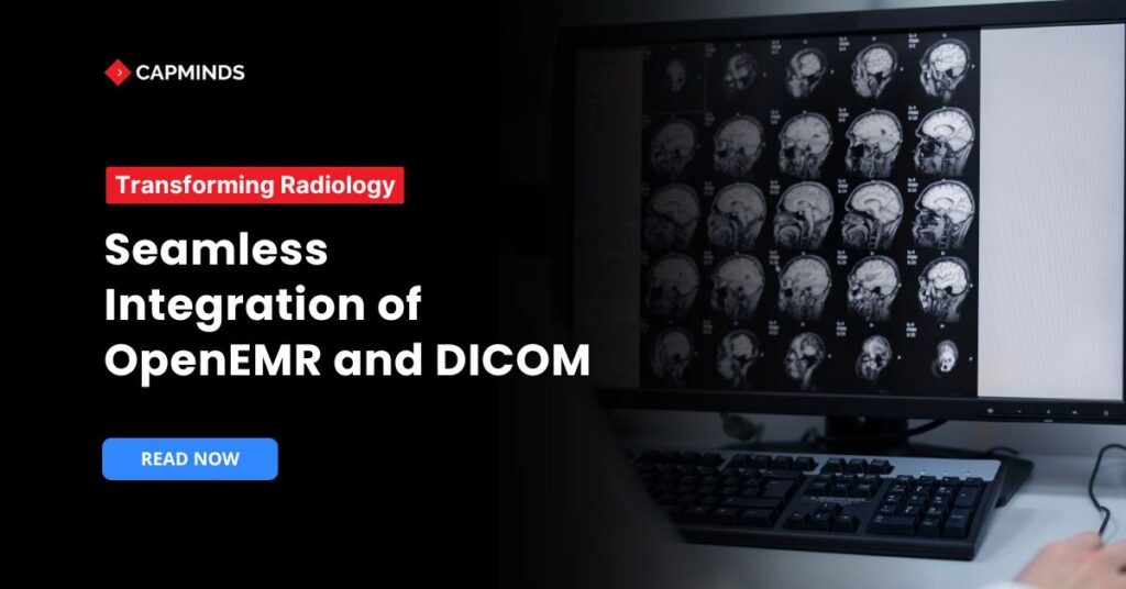
Radiology plays an important role in healthcare. It is involved in the diagnosis, treatment, and monitoring of health. With the development of technology, the incorporation of medical imaging with EMR systems is required to improve workflow efficiency and patient care.
Integrating DICOM with OpenEMR is still a challenge for many practitioners. In this blog, we’ll discuss the importance of DICOM and the steps on how to integrate OpenEMR with DICOM for a seamless radiology workflow.
What Is DICOM Integration in OpenEMR?
OpenEMR is an open-source software that is designed to help healthcare organisations maintain their electronic medical records. OpenEMR can be customised according to healthcare needs. It offers users to have full data ownership.
- OpenEMR is installed in over 15,000 healthcare centres globally.
- Medical Providers over 100,000 use OpenEMR and serve more than 200 million patients through their practice.
- OpenEMR is used in 100 countries all over the world and has been translated into 34 languages.
A DICOM Viewer is used to view, store, and manipulate medical images. Digital Imaging and Communications in Medicine is a standardized format for exchanging medical images.
- DICOM Viewer offers 3D reconstruction, enables anatomical views, and enhances images.
- It uses tools like distance, angles and areas for measuring images.
- DICOM allows creating reports, adding annotations, and sharing with professionals to get suggestions.
How DICOM Files, PACS, and EMRs Work Together
When a CT, MRI, or ultrasound scanner takes a picture, the scanner saves it in the DICOM file format, which includes both pixel data and metadata (patient name, modality, timestamps, and so on).
DICOM is vendor-neutral, enabling machines from Siemens, GE, Philips, and portable ultrasound devices to communicate in the same language. After acquisition:
- DICOM files are sent to a PACS server. A PACS is a dedicated archive for storing and managing medical images. It provides secure, economical storage and fast retrieval of images from multiple imaging modalities. PACS uses DICOM as its universal format, which means any compliant device or viewer can retrieve studies.
- The EMR references those images. The EMR receives HL7 or FHIR messages with study identifiers so it can link the imaging study to the patient chart. The images themselves remain in the PACS, but the EMR provides context, orders, reports, and lab results alongside the image.
- Clinicians access images through a DICOM viewer integrated into the EMR. When a clinician opens the patient record, the EMR calls the PACS, retrieves the relevant DICOM files, and displays them using a viewer. This process keeps patient data centralized and eliminates the need to log into multiple systems.
This integration reduces the delays and manual data entry associated with paper records or stand‑alone imaging systems. Centralized patient records and instant access to medical images support quicker diagnoses and better clinical decisions.
Secure DICOM PACS integration also helps organizations meet HIPAA and GDPR requirements and reduces the long‑term cost of imaging by using open‑source tools.
Related: The Beginners Guide to Using DICOM Viewer in OpenEMR
Benefits of DICOM Integration in OpenEMR for Radiology Practices
DICOM integration into OpenEMR assists in numerous ways.
1. Centralized Patient Records
DICOM integration in OpenEMR allows healthcare providers to access medical images directly within the EMR system without switching to different applications.
2. Workflow Efficiency
Through easy access to medical images, it reduces the delay in diagnosing and treatment plans. It allows providers to streamline the workflow and reduce manual data entry.
3. Diagnosis Capabilities
Integrating DICOM helps physicians and radiologists analyse high-quality medical images with patients records, lab results, respectively. This supports an accurate, faster diagnosis and better clinical decisions.
4. Image Storage and Retrieval
Medical images are stored in Picture Archiving and Communication in Medicine, which enables secure data encryption and authentication for access. It ensures safe, long-term storage of data and prevents unauthorized access.
5. Security Standards
DICOM is an interoperability standard for medical imaging. OpenEMR ensures security standards, regulations like HIPAA, GDPR, and other data protection laws.
6. Cost-Efficient
Integration of DICOM can be used in small clinics, hospitals to upgrade advanced medical imaging without spending on third-party solutions.
Challenges of OpenEMR DICOM Integration
- Without seamless integration, it might be a time-consuming task where users must upload, retrieve files manually. Uploading medical image data slows down processes.
- Due to fragmented patient records, images are stored separately, making diagnosis and treatment plans difficult.
- Providers may have difficulty viewing image results from different locations due to limited storage access.
- Compliance risks occur when security regulations are not enforced and controlled access to patient imaging data.
How OpenEMR Supports DICOM Integration
- While OpenEMR does not offer built-in DICOM support, third-party tools and modules allow seamless integration.
- OpenEMR can communicate with PACS servers that store and manage DICOM images.
- By adding web-enabled or standalone DICOM viewers, healthcare professionals can view medical images directly within OpenEMR.
- OpenEMR supports FHIR and HL7 standards, which gives it interoperability with radiology systems.
Related: The Comprehensive Features of DICOM Viewer Healthcare Practices Should Know
What Is a DICOM Viewer and Why Is It Important?
A DICOM viewer is specialised software that reads and displays DICOM files. It allows clinicians to view, manipulate, and annotate images on any computer without needing the original imaging equipment. In the context of an imaging EMR, the viewer is the bridge between the PACS and the clinician:
- DICOM viewers provide windowing, panning, zooming, measurement, and annotation tools that let radiologists and specialists examine images in detail. Alma Medical Imaging notes that while PACS digitises storage and distribution, it is the DICOM viewer that enables professionals to interpret and analyse images accurately.
- Because the viewer reads the standard DICOM format, images from any modality or vendor can be opened. The OpenEMR project points out that standardising image formatting through DICOM makes it possible for doctors to view and share images on any computer operating system.
- With an embedded viewer, clinicians can see imaging studies directly in the EMR instead of opening a separate application. This reduces context switching and facilitates remote consultations. Centralising images also helps reduce duplicate studies, which lowers radiation exposure and costs.
Modern DICOM viewers range from lightweight web‑based viewers to advanced workstations that support 3‑D reconstructions. In OpenEMR, the DICOM Web Viewer provides basic viewing and annotation capabilities; it is designed for review rather than diagnostic reporting.
For diagnostic reads, practices often integrate OpenEMR with enterprise PACS viewers or use certified third‑party viewers such as Weasis, RadiAnt or Horos.
DICOM Viewer vs PACS: Understanding the Difference
Both DICOM viewers and PACS work with medical images, yet they serve different purposes. The key differences are summarised below:
|
Component |
Primary role |
Key features |
|
DICOM Viewer |
An application that opens DICOM files and displays images for viewing and annotation |
Provides tools for window/level adjustments, zoom, measurement, and multi‑planar reconstruction; can be standalone or built into an EMR; not necessarily designed for long‑term storage |
|
PACS (Picture Archiving and Communication System) |
A server or service that stores, manages, and distributes imaging studies |
Centralised archive for DICOM files; manages storage lifecycle, user access and routing; often includes or integrates with a viewer; communicates with modalities via DICOM messages. |
Steps to Integrate DICOM with OpenEMR
Step 1: Setting Up OpenEMR for Imaging
Install OpenEMR on a secure server, configure necessary modules, and enable permissions for external integrations.
Step 2: Selecting a PACS Solution
Select a compatible PACS server like Orthanc that meets your organisation’s DICOM communication requirements.
Step 3: Configuring OpenEMR to Communicate with PACS
Install the selected PACS server and configure it to establish a DICOM connection between OpenEMR and the PACS.
Define DICOM nodes and ports to enable seamless data exchange. Test with sample patient images to ensure proper retrieval and display.
Step 4: DICOM Integration in OpenEMR
Select a DICOM viewer compatible with OpenEMR (e.g., Weasis, OsiriX, RadiAnt).
Configure OpenEMR to launch the viewer for image display. Test access to imaging studies directly from patient records.
Step 5: Ensuring Security Compliance
Implement user authentication and role-based access controls. Encrypt DICOM data transfers to meet HIPAA and GDPR.
Regularly audit and monitor system activity to ensure security compliance.
Future Trends in OpenEMR, PACS, and DICOM Integration
- Machine learning tools can assist radiologists in detecting abnormalities using AI-driven radiology diagnostics.
- Future PACS solutions will offer secure, scalable, and remotely accessible imaging storage, improving efficiency for healthcare providers.
- FHIR & HL7 advancements strengthen interoperability with radiology software systems.
As technology starts to advance, the integration of OpenEMR and DICOM is vital to improve diagnostic accuracy, patient care, and radiology workflows.
Seamless integration of OpenEMR and DICOM revolutionizes radiology workflows to ensure that healthcare professionals can store, retrieve, and analyze imaging data easily.
Using open-source PACS solutions and DICOM viewers, OpenEMR can be an efficient, cost-saving EHR for radiology clinics.
DICOM Viewer Customization Service by CapMinds
Want to get the most from the DICOM Viewer from your EMR systems? CapMinds is here to help you.
We are a professional health tech company with years of experience in EHR, EMR, OpenEMR, HL7 FHIR, Mirth Connect, Health Interoperability, and More. Our DICOM customization service includes:
- Custom Layouts and Toolbars
- Advanced Image Processing Capabilities
- Comprehensive Data Visualization
Our team of experts will closely work with you to tailor every aspect of the DICOM viewer in your EMR system, ensuring a smooth workflow.
Elevate your diagnostic capabilities and streamline your radiology practice with a DICOM viewer that truly puts you in control.
Contact CapMinds today and experience the full potential of the DICOM viewer for your Practice.
Frequently Asked Questions
Does OpenEMR include a built‑in DICOM viewer?
Yes. Since OpenEMR 5.0.4, the system includes an embedded DICOM Web Viewer developed by Jerry Padgett. This tool allows clinicians to upload, store, and view digital radiology images from within OpenEMR without installing additional software.
The viewer is designed for basic review and annotation, not for primary diagnostic reads. For diagnostic viewing, practices may need to integrate OpenEMR with a certified PACS viewer.
How do I open DCM files in OpenEMR?
OpenEMR offers two ways to view DICOM (.dcm) files:
- From the patient’s Documents tree. Upload the DCM file (or a zip of a DICOM study) into a folder under the patient’s Documents section. Clicking on the file will launch the DICOM Web Viewer. This method is useful for files stored on the server but can be slow if network bandwidth is limited.
- From the main menu. Navigate to Miscellaneous → DICOM Viewer and open the local DICOM file or folder using the file chooser. Opening files locally offers faster loading and is preferred for large studies. Once the study loads, use the toolbar to pan, zoom, and adjust window/level settings.
What is the difference between a DICOM viewer and PACS?
A DICOM viewer is software that displays images; it provides tools for interacting with them (zooming, measuring, annotating). A PACS is a storage and management system that archives imaging studies and handles DICOM communications. The viewer shows you the images, whereas the PACS securely stores them and manages access. As Stack Overflow users note, “The viewer is an application that allows you to look at images. The PACS is a server that stores the images”.
Is OpenEMR suitable for radiology practices?
OpenEMR can be adapted for radiology workflows but usually requires integration with imaging tools. The project’s DICOM Web Viewer allows basic viewing and annotation of DICOM studies, and OpenEMR can communicate with PACS servers and third‑party DICOM viewers. Practices needing advanced features (3‑D reconstructions, diagnostic reporting, Voice Recognition) often deploy a dedicated PACS with an FDA‑certified viewer while using OpenEMR for orders, scheduling and reporting.
Thanks to support for FHIR and HL7 standards, OpenEMR can exchange imaging orders and results with radiology information systems and imaging modalities.
How does EHR imaging integration improve patient care?
Integrating imaging into an EMR creates a single source of truth for patient data. In CapMinds’ experience, centralised patient records eliminate the need to log into separate systems and reduce duplicate studies. Workflow efficiency improves because providers can access images immediately when reviewing a chart.
Combining high‑quality images with lab results and clinical notes enables more accurate and timely diagnoses. A PACS also ensures secure, long‑term storage of imaging studies with proper encryption and authentication, supporting HIPAA and GDPR compliance.